64-Slice CT Scanning

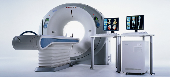
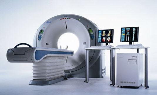
St. Mary’s state-of-the-art Toshiba Aquilion 64 CFX scanner is one of the fastest, most powerful computed tomography (CT) scanners on the market. Located right next to our Emergency Department for quick access when every minute matters, this scanner can synchronize with the patient’s heart rhythm and capture images between beats, offering high-speed, life-saving diagnostics.
“For the first time in medical history, there’s a technology fast enough to create images of the beating heart,” says Jeff Brown, St. Mary’s Director of Radiology and Cardiology Services. “The detailed images created by the 64-slice scanner make it possible for physicians to spot things that can’t be seen in images from older scanners, including the narrowing of artery walls that can cause a heart attack.”
“The scanner gives physicians the ability to detect disease early, while it’s most effectively treated,” Brown says. With early diagnosis, many patients are able to receive less invasive treatments with better outcomes than are possible with later diagnosis.
How it works
The Aquilion 64 is a vital tool in diagnosing a wide range of conditions, including heart attack and stroke. In just 15 seconds or so, the Aquilion 64 can create photo-quality images of the inside of the body — painlessly. It generates three-dimensional images of the heart, brain, circulatory system, skeletal system and internal organs, allowing physicians to diagnose narrowed arteries, internal bleeding, early stage cancers and much more.
Computerized tomography – CT – uses low-dose x-rays to create images of the inside of the body. Lying comfortably on the table, the patient holds his or her breath for a few seconds while the table slides through the gantry. As it does, the Aquilion silently takes 64 cross-sectional images – “slices” – every 400 milliseconds.
The Aquilion’s computer system quickly assembles thousands of these slices into 3-D images. The physician can look at these color images from any angle, isolate any organ, and zoom in on any spot. With the click of a mouse, the physician can look even deeper, seeing the chambers of the heart or the inside of arteries.
More than heart studies
“The Aquilion’s ability to conduct fast, highly detailed brain scans can help stop a stroke in its tracks,” Brown notes, “When a patient comes to St. Mary’s with stroke symptoms, the 64-slice CT scanner lets physicians determine within minutes if the patient can receive tPA.” TPA is a clot-busting medicine that can restore blood flow and prevent brain damage, but only stroke patients without bleeding in the brain can safely receive it. St. Mary’s Aquilion 64 has helped many patients safely receive tPA and reduce — and in some cases completely reverse — the effects of their stroke.
“It’s often difficult for children, or people with severe breathing problems, to hold their breath and remain still for the longer times needed by older scanners,” says Brown, “With the 64-slice, if they can hold still for just a few seconds, that’s all we need.”
The bottom line, Brown says, is that the Aquilion 64 makes better care possible.
“At the end of the day, what matters most is that we help patients prevent illness or recover faster and more completely,” Brown says.

St. Mary’s Toshiba Aquilion 64 CFX scanner, the only 64-slice scanner in the Athens area available for emergency diagnostics, provides highly detailed images in seconds. Only 64-slice CT technology is certified for studies of the heart and coronary arteries.

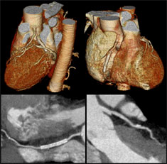
This scan, provided by Toshiba, shows a stent in a coronary artery in a view of the exterior of the heart (top) and also in cross-section through the coronary artery in which it was inserted (bottom).

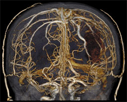
This scan, which took only 4 seconds, shows the blood vessels inside the brain. Such detail helps physicians diagnose strokes, aneurysms (including one visible on the right side of this image), brain tumors and more.
